“Every living cell tells a story — and learning to count them accurately is the first step in understanding life itself.”
Introduction: Why Cell Counting Matters
In every biological experiment — whether you’re growing bacteria, culturing mammalian cells, or analyzing blood — knowing how many cells you have is essential.
Cell counting helps researchers measure cell density, viability, and growth rate, forming the foundation for accurate experiments in microbiology, cell biology, and biomedical research.
And the best part? You don’t need a fancy machine.
You can count cells manually using a hemocytometer — a beautifully simple, yet powerful tool invented in the 19th century by Louis-Charles Malassez to count blood cells.
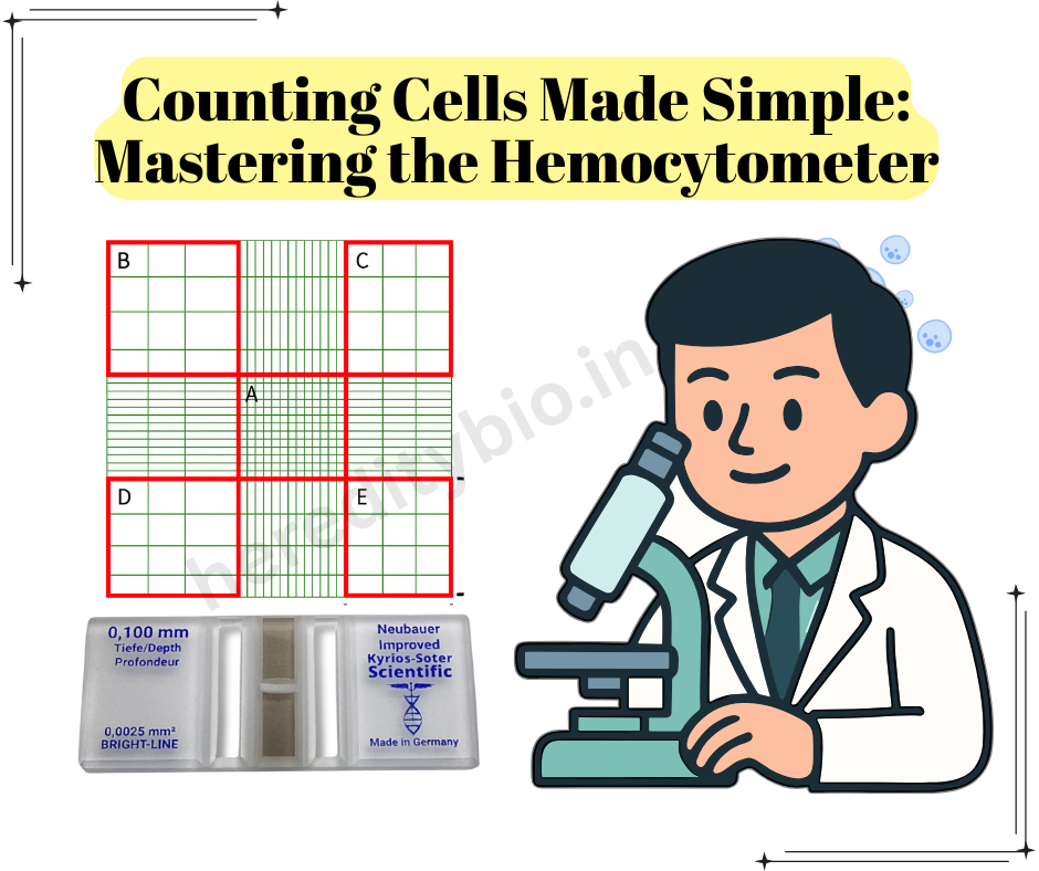
Meet the Hemocytometer
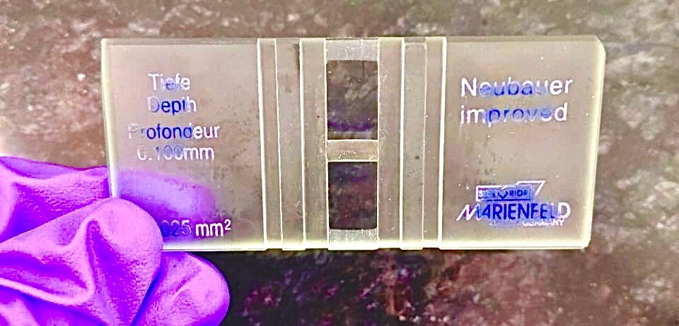
A hemocytometer is a thick glass slide with a tiny etched grid in the center.
When used with a coverslip, the grid holds a precise volume of liquid.
By counting the cells within certain squares, you can calculate how many cells are present per milliliter of sample.
Pro tip:
The etched grid ensures accuracy — each square is 1 mm × 1 mm, and the chamber depth is 0.1 mm, giving a volume of 0.0001 mL (100 nL) per square.
Learning by Doing: Step-by-Step Protocol
Here’s a simple 4-step guide that students can follow to confidently perform a cell count in the laboratory.
Step 1: Staining and Dilution
Goal: Distinguish live cells from dead ones.
Use Trypan Blue, a vital dye that only stains dead cells blue.
Mix your sample and dye in a 1:1 ratio (equal volumes).
- Example: 10 µL cell suspension + 10 µL Trypan Blue
- Note your dilution factor (2) — you’ll need it for your calculation later!
Step 2: Loading the Hemocytometer
Goal: Properly load the chamber to avoid bubbles or uneven cell distribution.
- Clean the hemocytometer and special thick coverslip with lens paper.
- Place the coverslip over the counting grid.
- Using a pipette, gently add 10 µL of the stained cell suspension into one of the V-shaped wells.
- The liquid fills by capillary action under the coverslip.
- Load the second chamber if you want to repeat or duplicate your count.
Tip: Don’t overfill — too much liquid will spill and distort the grid.
Step 3: Counting the Cells
Goal: Get an accurate and reproducible count.
- Place the hemocytometer on the microscope stage and focus on the grid using 10× objective.
- Count cells in five large squares — the four corners and the center.
- Follow the “top-right rule”:
- Count cells touching the top and right boundary lines.
- Skip cells touching the bottom or left boundaries (to avoid double-counting).
- Only count unstained (live) cells.
Tip: Aim for ~100 total cells for statistical accuracy. If the field is too crowded, dilute your sample further.

Step 4: Calculate Cell Concentration
Goal: Convert your counts into cells per mL.
Use the formula: Cells/mL=

Example:
- Total counted = 325 cells
- Dilution = 2 (1:1 Trypan Blue)
- Squares counted = 5

If your total sample volume is 5 mL, the total number of cells =
1.3×106×5=6.5×1061.3 \times 10^6 \times 5 = 6.5 \times 10^61.3×106×5=6.5×10⁶ cells.
Visualizing Your Data
Plot your counts in a simple table:
| Square | Cells Counted |
|---|---|
| Top Left | 62 |
| Top Right | 64 |
| Bottom Left | 63 |
| Bottom Right | 68 |
| Center | 68 |
| Total | 325 |
Average the counts from both chambers if you used both sides of the hemocytometer for better accuracy.
Student Insights & Common Mistakes
Do clean your coverslip and ensure capillary loading (no bubbles).
Do count evenly distributed fields — discard uneven samples.
Don’t forget to multiply by the dilution factor.
Don’t count dead (blue) cells in your viable cell count.
Why It Matters
Cell counting isn’t just a lab routine — it’s the first step in understanding cell growth, drug response, and health.
Whether you’re testing a cancer therapy, culturing stem cells, or studying microbial growth, your results depend on accurate and reproducible counts.
Learning this technique early helps students:
- Strengthen observation and quantitative skills
- Develop accuracy and patience
- Understand how small-scale data translates into biological meaning
Conclusion
Using a hemocytometer is one of the most empowering and fundamental skills in life sciences.
It teaches precision, care, and scientific curiosity — qualities that define great scientists.
So, next time you look through that microscope, remember:
Each cell you count is a tiny universe of life, waiting to be understood. 🌿
#hemocytometermastery #cellcounting #cellculture #laboratorytechniques #lifescience #bioresearch #cellbiology #microscopy #lablife #scienceeducation #researchskills #biotech #scientistlife